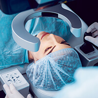
Recently, Dr. Gollamudi was featured in Cataract and Refractive Surgery Today, about his thoughts and experiences with laser cataract surgery. Take a look at his article below!
Capitalizing on technology allows doctors to take our oath to “do no harm” to a higher level. One good technology plus another good technology creates a synergy that we all strive for – a safer, more effective surgery with lower
downside potential. By marrying technologies, we create a safety zone, increasing our confidence and ability to minimize complications, even in the most difficult cases.
CASE 1
A 67-year-old woman presented with congenital zonular dehiscence, which under a widely dilated pupil indicated a visibly “scalloped” edge along 3 to 4 clock hours. The lens also shifted to the other side and lacked adequate zonular support. The patient’s presurgical BCVA was 20/50. Using the Catalys Precision femtosecond laser (Abbott Medical Optics), centration of the capsulotomy was automatically calculated. When the treatment was done, it appeared off-center; however, once the capsular tension ring was inserted, a perfectly centered capsulotomy was apparent. This critical step was rendered far simpler because this laser system has the ability to calculate placement despite the initial
decentration of the patient’s lens. Next, the surgeon used the laser to perform complete segmentation and softening of the nucleus, which substantially reduced manipulation during lens removal and helped prevent damage to any additional zones. Phacoemulsification using the venturi pump mode on the WhiteStar Signature (Abbott Medical Optics) allowed nuclear fragments to flow easily into the phaco tip. The ability to flip on the fly from peristaltic to venturi pump mode permitted a safe distance between the phaco probe and the fragile capsule, thus minimizing risk caused by stress on, tension on, or manipulation of the capsule. The surgeon inserted a capsular tension ring to stretch the lens capsule
into an ideally shaped, round, centered capsule allowing for implantation and a perfectly centered IOL. The patient had a postoperative BCVA of 20/30, and no new zonular damage was caused. The pairing of these technologies produced a great outcome for a case carrying a high risk for capsular rupture or the inability to achieve IOL cent ration.
CASE 2
A 73-year-old man presenting with a dense cataract and count fingers at 1 foot BCVA in his right eye underwent surgery using the femtosecond laser to fragment the nuclei. Laser fragmentation contributes to the safety of the procedure by reducing or even eliminating the amount of ultrasound energy required to carve, crack, and disassemble the nucleus. A 20-gauge MST/Dewey large-bore phaco tip (30º bend, 0.7-mm diameter, and 0.9-mm outer diameter; MicroSurgical Technology) was used to complement the fluidics of the WhiteStar. By combining the larger-bore tip with the venturi pump mode, the fragments were drawn out and removed without expending any extra phaco energy. The larger internal bore of the phaco needle allowed larger nuclear fragments to be removed without the need for further ultrasound fragmentation. The patient’s postoperative UCVA was 20/25. In cases involving dense nuclei, emulsification can prove difficult, posing increased risk for eye trauma. These cases carry a much higher risk of pseudophakic bullous keratopathy from endothelial damage and capsular rupture, necessitating some other form of securing the lens, either in the sulcus with a posterior chamber fixated lens or even an anterior chamber IOL. By pairing technologies, the surgeon maximizes the benefits and creates a safer case by preserving the endothelium and zonules, avoiding complications caused by manipulation, and reducing exposure to phaco energy.
LIMITATIONS
There are, of course, limitations with any technology. The biggest hurdles to incorporating femtosecond laser-assisted cataract surgery in a practice are expense and financing. This state-of-the art equipment is a costly investment, although many companies now offer extended payment options or rental programs. Recouping the investment can take time, particularly because treatment with femtosecond laser-assisted cataract surgery is not covered by insurance, and many patients simply do not have the extra income to take advantage of the procedure. Laser cataract surgery is not an option for all patients—even for those are willing to pay for it. The laser has to dock with the eye near the limbus. Docking may be prevented (1) because the palpebral fissure is too small, (2) there is a prior surgical bleb from glaucoma surgery or a previously placed seton, or (3) scar tissue prevents transmission of the laser energy into the eye. The laser only transmits through clear tissue; so patients who present with corneal scarring may not be ideal femtosecond laser candidates. The laser treatment creates penetrating arcuate incisions in the cornea that produce gas, which could track along the prior incisions and open them. Patients who have had a corneal transplant or scarring from a radial keratotomy that could block the laser must be carefully evaluated for candidacy.
IN SUMMARY
Cataract surgery has continually improved, with new techniques, instruments, and technology. Combining some of the newest innovations can create synergy, allowing for a safer and more predictable outcome. In my hands, use of a different phaco tip, in combination with femtosecond laser treatment and a variety of settings on the ultrasound/irrigation/aspiration apparatus, has allowed me to better handle both routine and difficult cases. As exemplified by the two cases I reported, surgeries that previously could turn into “nightmares” are now being completed without difficulty. I look forward to further changes in techniques, as the ophthalmic community pushes forward with new technology.


